Lipid bilayer: Difference between revisions
| Line 9: | Line 9: | ||
==Confusion with other twos== | ==Confusion with other twos== | ||
===Two-tailed | ===Two-tailed phospholipids=== | ||
A phospholipid is a ''two-tailed'' lipid. Thus, a phospholipid bilayer has two ''two''s in it: one describing the two tails of each molecule, and the other describing the fact that the layer is two molecules thick. The ''bi''layer refers to the latter. Note that whereas the two tails of each molecule are (approximately) parallel to one another, so that their thickness doesn't add up, the thickness of the two layers of the bilayer does add up (because they stack up on top of each other). | A phospholipid is a ''two-tailed'' lipid. Thus, a phospholipid bilayer has two ''two''s in it: one describing the two tails of each molecule, and the other describing the fact that the layer is two molecules thick. The ''bi''layer refers to the latter. Note that whereas the two tails of each molecule are (approximately) parallel to one another, so that their thickness doesn't add up, the thickness of the two layers of the bilayer does add up (because they stack up on top of each other). | ||
Latest revision as of 08:40, 15 December 2024
Definition
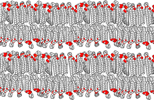
A lipid bilayer is a thin membrane (surrounding a cell or organelle) that is two molecules thick, with both molecules being lipids with a hydrophilic head and hydrophobic tail. The hydrophilic head of the outer molecule points outward to the (usually aqueous) external environment, and the hydrophilic head of the inner molecule points inward to the cell or organelle being surrounded by the bilayer. The hydrophobic tails both point inward toward each other.
Lipid bilayers constitute biological membranes, which, in addition to the lipid bilayer, contain embedded proteins called integral membrane proteins. Examples of biological membranes are cell membranes (in both prokaryotic cells and eukaryotic cells) as well as the membranes of cellular organelles (mostly in eukaryotic cells). The most typical examples of lipid bilayers that occur in cell membranes and organelle membranes are those where the lipid is a phospholipid. Other possibilities for the lipid include sphingolipids, glycolipids, and cholesterol.
Confusion with other twos
Two-tailed phospholipids
A phospholipid is a two-tailed lipid. Thus, a phospholipid bilayer has two twos in it: one describing the two tails of each molecule, and the other describing the fact that the layer is two molecules thick. The bilayer refers to the latter. Note that whereas the two tails of each molecule are (approximately) parallel to one another, so that their thickness doesn't add up, the thickness of the two layers of the bilayer does add up (because they stack up on top of each other).
Double bilayers
There are some contexts where biological membranes are double membranes:
- In the case of the nucleus, the membrane is folded on top of itself
- In the case of the mitochondrion and chloroplast, there is a distinct inner mitochondrial membrane and outer mitochondrial membrane)
The double here is distinct from the bilayer double. In the case of a double membrane where each membrane constitutes a bilayer, we expect a -molecule thick layer. Note that for organelles with distinct inner and outer membranes, there is an intermembrane space (such as intermembrane space of mitochondrion) whose thickness is a small multiple (within an order of magnitude) of the thickness of the lipid bilayers.
Summary
| Item | Value |
|---|---|
| Type of structures that have lipid bilayers as their membranes | both prokaryotic cells and eukaryotic cells have cell membranes that are made of lipid bilayers. In addition, the organelles in eukaryotic cells, such as the nucleus, mitochondrion, lysosomes, and others, have their own membranes which comprise lipid bilayers. |
| Size | Thickness: The bilayer is only two molecules thick, so its thickness is a few nanometers (nm), where a nanometer is meters. |
| Chemical constituents | The lipid molecules, which are usually phospholipids in the case of cell membranes and organelle membranes. Note that the lipid making up the outer leaflet may differ from the lipid making up the inner leaflet. |
| Function | Controls the entry and exit of materials between the cell or organelle and the environment. Specifically, the hydrophobic tails attempt to block water-soluble substances from crossing the lipid bilayer. There are a number of special mechanisms in use (depending on the cell or organelle) that are used to transport materials across the membrane. |
Size
Thickness
The bilayer is only two molecules thick, so its thickness is a few nanometers (nm), where a nanometer is meters.
The exact thickness depends on the specific choice of the lipid, and in particular the length of its hydrophobic tail.
Comparison with cell sizes
Comparison with prokaryotic cells: Prokaryotic cells have diameters in the 1000-10000 nm range, which is about 100-1000 times the thickness of the lipid bilayer.
Comparison with eukaryotic cells: Eukaryotic cells have diameters in the 10000-1000000 nm range, which is about 1000-10000 times the thickness of the lipid bilayer.
Comparison with wavelengths of light and implication for visibility under microscopes
The thickness of the lipid bilayer is considerably smaller than the wavelength of visible light (400-700 nm). Light microscopes have resolutions limited to about 200nm, and hence cannot be used to study these bilayers. Lipid bilayers can be studied using electron microscopes or fluorescence microscopes. However, the study of these bilayers is quite difficult due to their fragility.
Physical structure
Outer leaflet
The outer leaflet refers to the outer half of the bilayer which has a hydrophilic head pointing to the aqueous external environment and a hydrophobic tail pointing inward and facing the hydrophobic tail of the inner leaflet.
Note that the "tail" here refers to all the hydrophobic tails of the molecule. If the molecule is two-tailed (as is the case with phospholipids) the two tails both point in the same direction, toward the inner leaflet. If the molecule is three-tailed, the three tails point in the same direction, toward the inner leaflet.
Inner leaflet
The inner leaflet refers to the inner half of the bilayer which has a hydrophilic head pointing to the aqueous environment of the enclosed cell or organelle and a hydrophobic tail pointing outward and facing the hydrophobic tail of the outer leaflet.
Note that the "tail" here refers to all the hydrophobic tails of the molecule. If the molecule is two-tailed (as is the case with phospholipids) the two tails both point in the same direction, toward the outer leaflet.
Chemical constituents
Hydrophilic head and hydrophilic tails
Note that this applies separately to each individual lipid molecule in the lipid bilayer (in either the outer leaflet or the inner leaflet). Different lipid molecules could be of different types, and (with the exception of cholesterol), even within the same type, there could be many different choices of actual molecules depending on the selection of fatty acid(s) and potentially the choice of hydrophilic head.
Below are some of the possibilities for the head and tails based on the kind of lipid.
| Type of lipid | Hydrophilic head | Type of tail | Number of tails |
|---|---|---|---|
| phospholipid | phosphate | fatty acid | 2 |
| sphingolipid | sphingoid base of some sort | fatty acid | 1 (at least, 1 usable, maybe 2 in theory?) |
| glycolipid | sugar residue | fatty acid | 1 |
| cholesterol | hydroxyl group | rest of the cholesterol molecule | 1 |
Selection of fatty acid and whether and how saturated it is
Each of the fatty acid tails could be chosen as a saturated or unsaturated fatty acid (here, "saturated" means only single C-C bonds; unsaturated means at least one C=C double bond). The unsaturated fatty acid could be chosen as a monounsaturated or polyunsaturated fatty acid. The polyunsaturated fatty acid could be chosen as an -3 or -6 fatty acid.
In general, greater use of unsaturated fatty acids results in greater flexibility of the biological membrane, and greater use of saturated fatty acids results in greater rigidity of the biological membrane. The mechanism is as follows: the presence of double bounds forces the fatty acid to take up more space, due to local rigidity created by the pi-bond. This means that the fatty acid tails cannot be packed that much, and have a bit more free space, and this is what gives the membrane flexibility. Conversely, with only single bonds, the tails can be packed more tightly. So, it seems that the greater local rigidity imposed by double bonds is what creates overall more flexibility for the membrane.
